The Weberian apparatus of the zebrafish, Danio rerio
Rebecca Jordan (2011)
The Weberian apparatus is a specialized auditory structure of the axial skeleton of otophysan fish (series Otophysi, within the superorder Ostariophysi). It comprises four bony elements or Weberian ossicles, derived from parts of the anterior-most vertebrae, which via their ligamentous connections form a chain linking the swimbladder to the inner ear. From posterior to anterior, the Weberian ossicles are the tripus, intercalarium, scaphium and claustrum. The tripus is connected via ligaments to the anterior portion of the swimbladder, while the anterior elements are coupled to the inner ear fluid space (the atrium of the sinus impar). The Weberian ossicles mechanically couple vibrations of the swimbladder induced by sound to the inner ear fluid, allowing sound to be transduced into electrical signals by the sensory cells there. The function of the Weberian ossicles is therefore in some respects analogous to that of the middle ear ossicles of terrestrial vertebrates, although these structures are completely unrelated.
There is considerable variability in the structure of the Weberian apparatus among the numerous species of otophysan fish (Ladich & Popper, 2004). We investigated the morphology of the zebrafish Weberian apparatus through micro-CT scanning and alizarin red staining: some of our results are shown as Figs. 1-3 below. In zebrafish, the tripus is the largest Weberian ossicle, connected via the very delicate intercalarium to the scaphium. The claustrum, located dorsal to the scaphium (Fig. 2), is described as being fused to the first vertebra and thus immobile in zebrafish, functioning to support the wall of the sinus impar atrium (Bang et al., 2001). Development of the Weberian apparatus as zebrafishes grow coincides with an expansion of maximum frequency detection from 200 to 4000 Hz, suggesting that these structures have a role in the detection of higher sound frequencies (Higgs et al., 2002). It is becoming clear as research progresses that the ears of fishes are much more specialized than previously believed.
 |
 |
 |
||
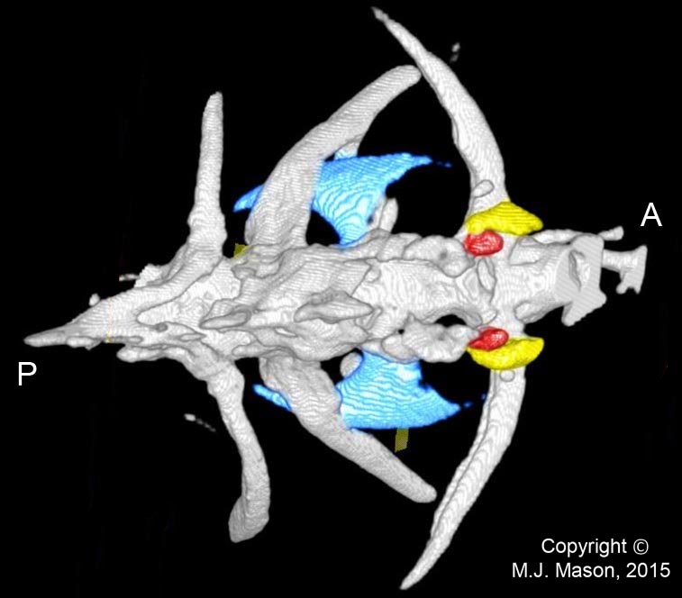
Fig. 1. Micro-CT image of the most anterior vertebrae of an adult male zebrafish, dorsal view. The tripus is coloured blue, the scaphium yellow and the claustrum red. A = anterior; P = posterior. The intercalarium, which is located between the scaphium and tripus, is not visible in this reconstruction, presumably due to scan resolution limitations. |
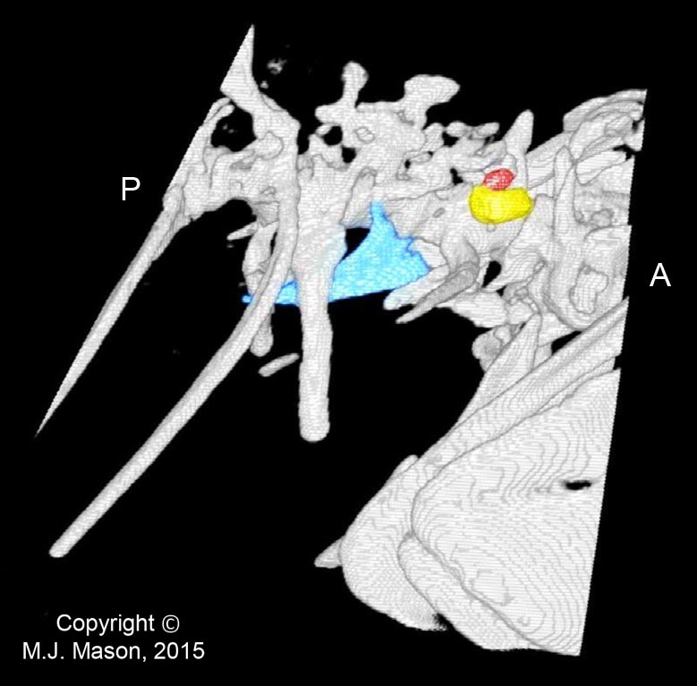
Fig. 2. Right lateral view of the same micro-CT reconstruction. |
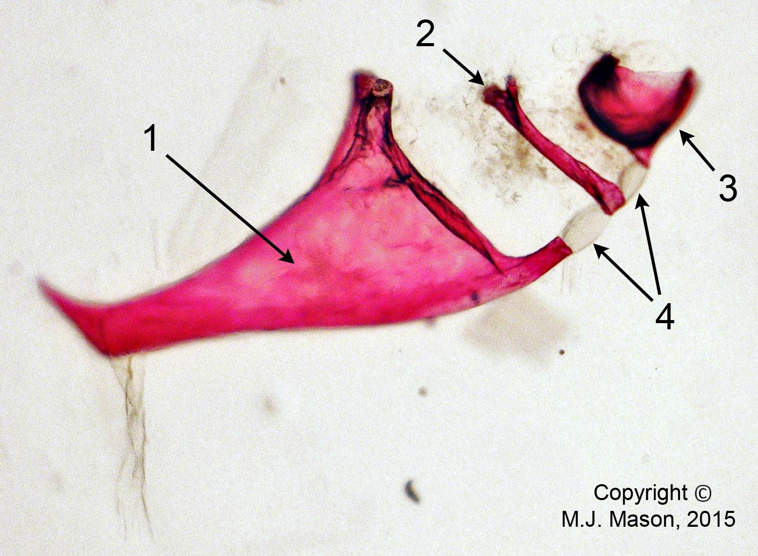
Fig. 3. Photomicrograph (taken at 40x magnification) of three of the four Weberian ossicles dissected from an adult male zebrafish. The tripus (1), intercalarium (2) and part of the scaphium (3) are still attached in their chain via inter-ossicular ligaments (4). The fish had been fixed with PFA and stained using the alizarin red staining process, rendering the flesh transparent and calcified matrix (such as bone) pink. |
References
Bang, P.I., Sewell, W.F. & Malicki, J.J. (2001) Morphology and cell type heterogeneities of the inner ear epithelia in adult and juvenile zebrafish (Danio rerio). Journal of Comparative Neurology 438: 173-90.
Higgs, D.M., Rollo, A.K., Souza, M.J. & Popper, A.N. (2003) Development of form and function in peripheral auditory structures of the zebrafish (Danio rerio). Journal of the Acoustical Society of America 113: 1145-54.
Ladich, F. & Popper, A.N. (2004) Parallel evolution in fish hearing organs. In: Evolution of the Vertebrate Auditory System (eds Manley, G.A., Popper, A.N. & Fay, R.R.), pp. 95-127. New York: Springer.
This work represents the results of a short undergraduate project performed in the Mason and Fleming laboratories by Rebecca Jordan in 2011.
Please contact Dr. Matthew Mason for more information about this as yet unpublished work.
With grateful thanks to Dr. Angeleen Fleming.